
Imaging Essentials: Small Animal Abdominal Ultrasonography – A Tour of the Abdomen: Part 2
As originally published in Today's Veterinary Practice," March/April 2016 (Volume 6, Number 2)
Danielle Mauragis, AS, CVT, and Clifford R. Berry, DVM, Diplomate ACVR
University of Florida
Welcome to our series of articles on small animal abdominal ultrasonography. The first 3 articles provided an overview of basic ultrasonography principles, while the previous article and this one review a systematic pattern for scanning the abdomen. Future articles will discuss scanning principles for each abdominal organ and system, normal sonographic appearance of abdominal organs and systems, and identification of common abnormalities seen during ultrasound examination.
Abdominal ultrasonography is a noninvasive technique that provides cross-sectional anatomy of the organs of the abdomen based on differences in acoustic impedance. The first 3 articles in this series, available at tvpjournal.com, have discussed:
- Basics of Ultrasound Transducers & Image Formation (January/February 2015)
- Physical Principles of Artifacts & False Assumptions (May/June 2015)
- Basics of Image Optimization—How to Obtain High-Quality Scans (November/December 2015).
ABDOMINAL TOUR
A systematic pattern for scanning the abdomen is an important aspect of any abdominal ultrasound (US) examination. In approaching abdominal ultrasonography, the practitioner should identify the questions the scan needs to answer; however, a negative US scan does not rule out disease.
The systematic approach:
- Starts in the cranial abdomen (at the liver)
- Proceeds in a clockwise fashion that extends around the outside of the abdomen
- Comes back in a counterclockwise fashion to incorporate the gastrointestinal tract and middle abdomen (see Checklist for Systematic Approach to the Abdomen, available at tvpjournal.com).
The first part of this article covered step 1 and part of step 2; this article discusses the remainder of step 2 and step 3 (Table). Part 1 also discussed:
- Basics of driving the transducer
- Probe orientation
- Identification of abnormalities.

PATIENT POSITIONING
For abdominal US, the dog or cat can be in dorsal or lateral recumbency; both scanning techniques are equally effective. This article series discusses a systematic approach to scanning the abdomen using a dorsally recumbent technique; however, the approach is just as applicable to scanning in lateral recumbency. The sonographer must learn to scan the patient in either position.
TOUR OF THE ABDOMEN
Left Medial Iliac & Hypogastric Lymph Nodes
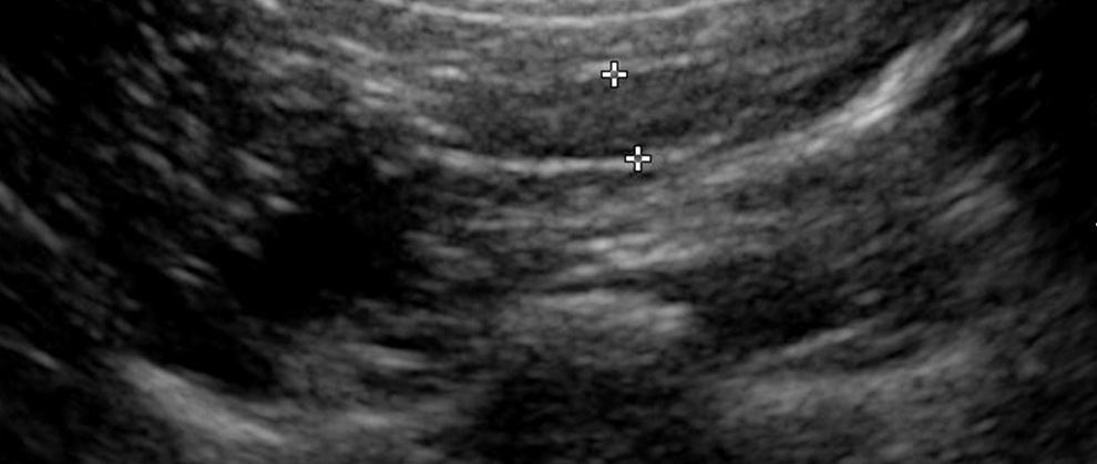
With the transducer in long axis, align the aorta in long axis at the level of the caudal abdominal aortic trifurcation into the left external iliac artery. The left medial iliac lymph node is seen as a hypoechoic fusiform- to oval-shaped structure located along the lateral (near field) aspect of the trifurcation (Figure 1).
The hypogastric lymph node is located between the right and left external iliac arteries, ventral to the continuation of the caudal abdominal aorta. Visibility of this lymph node in a normal dog and cat varies; however, because it is important to look for enlargement of this lymph node, knowing the correct anatomy is essential.

Figure 1. Long-axis image just lateral to the trifurcation of the caudal abdominal aorta, with a fusiform-shaped hypoechoic structure noted in the near field. This is the left medial iliac lymph node adjacent to the left external iliac artery, just after the caudal abdominal aortic trifurcation.
Colon, Uterine Body, & Trigone
If the patient is an intact female, rotate the transducer into transverse orientation at the level of the trigone of the urinary bladder and look for the colon, uterine body, and trigone (Figure 2). The uterine body can be traced caudally (distance motion) to the level of the cervix and cranially into the uterine horns.

Figure 2. Transverse image of the colon and uterus (arrow) in a cat. The uterus is a hypoechoic tubular structure located between the urinary bladder and colon, although the urinary bladder is not pictured in this image.
Right Medial Iliac Lymph Node
The right medial iliac lymph node is located lateral to the trifurcation of the caudal abdominal aorta and right external iliac artery.
Right Kidney & Right Adrenal Gland
With the transducer in long axis, trace the caudal vena cava along the right side of the abdomen to the level of the right kidney (Figure 3). Then evaluate the right kidney in long and short axes (Figure 4). With the transducer in long axis, angle it medial to the right kidney. The right adrenal gland is located next to the caudal vena cava (Figure 5).

Figure 3. Long-axis image of the caudal vena cava (near field) and the abdominal aorta (far field) from the right paralumbar position in a dog.

Figure 4. Long-axis sagittal plane view of the right kidney in a dog (A). Short-axis transverse plane view of the right kidney in the same dog (B). The renal hilum, renal pelvis, and vasculature are located centrally in the transverse image.

Figure 5. Long-axis image of the right adrenal gland (arrow) adjacent to the caudal vena cava at the level of the right kidney (not seen in this image) in a dog.
One can then reevaluate the right side of the liver and the gallbladder. The outer clockwise circle for the abdomen is complete.
Gastrointestinal Tract & Pancreas
In the midabdomen, place the transducer in long axis just caudal to the liver so that a transverse view of the stomach is seen. Slide the transducer caudal to identify the transverse colon in the transverse imaging plane (Figure 6). The left lobe of the pancreas is located between the stomach and the colon, adjacent to the splenic portal vessels that cross the abdomen from left to right to form the portal vein (Figure 7).

Figure 6. Long-axis image of the stomach (ST), spleen (SP), and colon (CO). This forms the splenic triangle and is the location of the pancreas. Often the pancreas is isoechoic to the fat in the mesentery and cannot be identified as a separate structure.

Figure 7. Left lobe of the pancreas with the central pancreatic duct and adjacent splenic portal vessels.
Rotate the transducer into the transverse imaging plane and slide it along the right side of the stomach to the pyloroduodenal junction (Figure 8). With the transducer in the transverse imaging plane (with the notch toward the right of the patient), follow the duodenum distally. The right lobe of the pancreas is seen along the mesenteric surface of the duodenum (Figure 9).

Figure 8. Transverse (short-axis) imaging plane of the pyloroduodenal junction in a dog (A) and a cat (B). In each case, the pylorus is on the right of the image and the duodenum is on the left. The thickening of the muscularis layer (arrow) is the pyloric sphincter.

Figure 9. Transverse (short-axis) imaging plane of the duodenum; note the right lobe of the pancreas along the mesenteric border of the duodenum. The ascending colon is just medial to the right lobe of the pancreas (right side of image).
Medial to the duodenum are the ascending colon and cecum. Along the medial surface of the ascending colon, look for the ileum as it comes into the ascending colon at the ileocolic junction (Figure 10). The differences in the bowel segments will be discussed in detail in a future article that focuses on the gastrointestinal tract.

Figure 10. Longitudinal image of the ileocolic junction (arrow) in a normal dog.
Adjacent to the ileocolic junction is a small collection of lymph nodes that drain this region (Figure 11).

Figure 11. Oblique imaging plane view of the lymph nodes (small oval hypoechoic structures) surrounding the ileocecocolic junction in a cat.
Midabdomen & Mesenteric Lymph Nodes
To scan the midabdomen, make sure the entire area is evaluated, which requires developing and using a repeatable method for ensuring coverage.
- Starting cranial and along the left, with the transducer in long axis at the level of the spleen, slide the transducer to the right across the abdomen to the level of the right kidney.
- Slide the transducer caudally, ensuring an overlap in the field of view.
- Now slide the transducer back to the left.
- Continue in this “mowing the lawn” fashion until you reach the urinary bladder trigone.
The mesenteric lymph nodes are located just to the right of the umbilicus at the level of the mesenteric portal vessels and branches of the cranial mesenteric artery (Figure 12).

Figure 12. Longitudinal axis (oblique sagittal) imaging plane of a normal mesenteric lymph node (arrow) in a dog.
Once at the urinary bladder trigone, rotate the transducer to the transverse imaging plane and slide it cranially along the descending colon.
If the dog is an intact male, you can evaluate the testicles by using a high-resolution, high-frequency transducer (Figure 13). This completes the abdominal tour.

Figure 13. Longitudinal axis of the left testicle in a dog. The echotexture is compact and uniformly hyperechoic. The bright echogenic line centrally represents the mediastinum testes.
ABDOMINAL TOUR TIME
The time it takes to complete the total abdominal scan depends on the type and size of the animal and the presence of any abnormalities. Scanning a normal cat abdomen may take only 10 minutes, while an abnormal scan on a large breed dog can take 45 minutes to 1 hour. Be sure to allocate enough time to perform the complete examination.
IN SUMMARY
A complete abdominal examination requires patience and time. A methodical approach to the abdomen is necessary so that all aspects of the abdomen are scanned with the appropriate images being obtained.
If the transducer is not placed over the area of abnormality, changes cannot be identified. Even when the transducer is placed over the abnormal area, if there are no differences in acoustic impedance, the abnormality will not be visualized as different from the normal structure or organ being evaluated.
Current survey abdominal radiographs must be obtained before ultrasound evaluation as ultrasonography is not a good survey tool for the abdomen. A negative scan does not rule out disease.
US = ultrasound
Suggested Reading
Kremkau F. Sonography Principles and Instruments, 8th ed. Philadelphia: Saunders Elsevier, 2010.
Mattoon J, Nyland T. Small Animal Abdominal Ultrasound, 3rd ed. Philadelphia: Saunders Elsevier, 2014.
Penninck D, d’Anjou M. Atlas of Small Animal Abdominal Ultrasound, 2nd ed. Ames, IA: Wiley Blackwell, 2015.

Danielle Mauragis, AS, CVT, is a radiology technician at University of Florida College of Veterinary Medicine, where she teaches diagnostic imaging. She coauthored the Handbook of Radiographic Positioning for Veterinary Technicians and received the Florida Veterinary Medical Association’s 2011 Certified Veterinary Technician of the Year award.

Clifford R. Berry, DVM, Diplomate ACVR, is a professor of diagnostic imaging at University of Florida College of Veterinary Medicine. His research interests include cross-sectional imaging of the thorax, nuclear medicine, and biomedical applications of imaging. He received his DVM from University of Florida and completed a radiology residency at University of California–Davis.
The Rubik's Cube is a three-dimensional twisty puzzle. Learn the easiest Rubik's Cube solution here.